Visual computing has developed sophisticated research approaches for radiation oncology. However, only a few have been fully integrated into daily clinical practice, patient care, and industrial products.
This online workshop series aims to:
![]() bridge specialists from the domains of visual computing and radiation oncology that are interested in collaborating on cutting-edge topics of research.
bridge specialists from the domains of visual computing and radiation oncology that are interested in collaborating on cutting-edge topics of research.
![]() provide a space for networking and interacting, for nurturing new ideas, and for initiating collaborations between the two domains.
provide a space for networking and interacting, for nurturing new ideas, and for initiating collaborations between the two domains.
![]() provide a better understanding of visual computing solutions (and novel trends in the field) that are applicable to research questions of radiation oncology.
provide a better understanding of visual computing solutions (and novel trends in the field) that are applicable to research questions of radiation oncology.
We target everyone that has an interest in how novel strategies from the domain of visual computing can be applied to radiation oncology research and practice. You are invited and encouraged to join—whether you work in academia, clinic or industry!
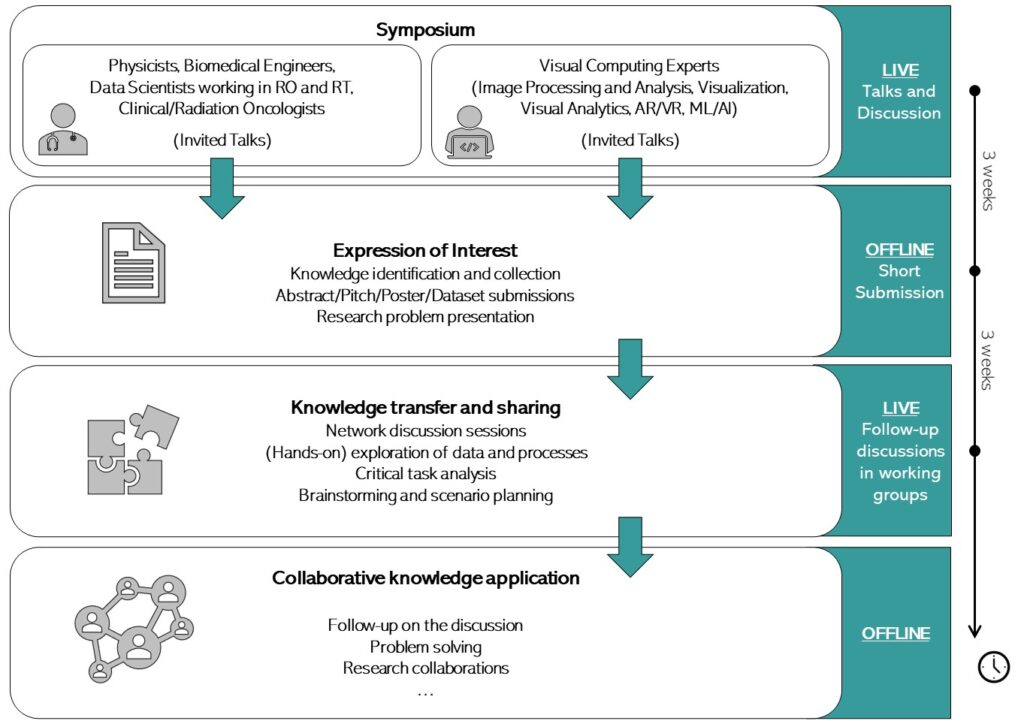
Format
First Thematic Session: “Multimodal and Multiparametric Approaches”
(Netherlands Cancer Institute)
Multi-parametric imaging for tumor characterization and target delineation
The delineation of a tumor as the target for irradiation is a key challenge in radiation oncology. Usually, images from multiple sources, such as CT, PET and MRI are used. For example, in prostate cancer the tumor inside the prostate is delineated based on a combination of anatomical images (T2-weighted MRI) and images reflecting diffusion of water in and between cells (Diffusion-weighted MRI) and blood perfusion of the tissue (Dynamic contrast-enhanced MRI). For clinicians, the challenge is how to combine these images in such a way that optimal choices for delineation are made. For researchers the challenge is to identify how these images reflect histological characteristics of the tumor. In this presentation results from a multi-center collaboration, validating multi-parametric MRI with histopathology of prostate specimen, will be shown.
Short Bio: Uulke van der Heide received his training as medical physicist at the department of radiotherapy of the University Medical Center in Utrecht, the Netherlands and worked there as a medical physicist until 2011. He now works as a medical physicist and group leader at the Netherlands Cancer Institute in Amsterdam, the Netherlands. He holds a chair as professor of imaging in radiotherapy at the Leiden University Medical Center. He participates as teacher and course director in the ESTRO school. His research group works on the improvement of target definition in radiotherapy by application of MRI and the development and validation of quantitative imaging methods for tumor characterization for radiotherapy dose painting. He further leads the MR-guided radiotherapy program at the Netherlands Cancer Institute.

University of Bergen &
Haukeland University Hospital
Multi-modal medical visualization
Advances in medical imaging techniques are bringing more and more different imaging modalities that provide additional information on anatomy and physiology. For instance, a single patient can have a CT scan, PET scan, as well as an MRI scan with different weighted images. When there is more than one modality acquired, mental integration of the different contrasts between the different images becomes more challenging. Our research team aspires to develop novel interactive visualization approaches for improved exploration, analysis, and communication of such multimodal medical imaging data. Our current focus in this context is on multi-parametric MR acquisitions. In this talk, I will provide a general introduction to medical visualization and highlight several visualization applications aimed at improved visual analysis for multimodal imaging data related to ongoing gynecological cancer and multiple sclerosis research.
Short Bio: Noeska Smit is an Associate Professor (tenure track) in the visualization research group at the University of Bergen, Norway, since 2017, where she leads a team researching multimodal medical visualization. She is also affiliated to the Mohn Medical Imaging and Visualization (MMIV) centre as a senior researcher at the Haukeland University Hospital. After working as a radiographer for three years, she completed her studies in Computer Science at the Delft University of Technology, the Netherlands, specializing in Computer Graphics and Visualization in 2012. In 2016, she obtained her PhD in medical visualization at the same institute in collaboration with the Anatomy and Embryology department at the LUMC in Leiden. Currently, she is researching novel interactive visualization approaches for multimodal medical imaging data. Her current focus in this context is on multi-parametric MR acquisitions.
1. Symposium (7 April 2021, 14.00-16.00 CET)
The symposium will feature invited thematic talks from two specialists: Uulke van der Heide (medical physics) and Noeska Smit (medical visualization), followed by a first round of discussions and interactions. To register, use this link. You will receive a participation link for ZOOM a few days before the symposium.
2. Expression of Interest (28 April 2021, by 23.00 CET)
Participants can express their interest in continuing the discussions, by submitting abstracts, posters, pitches, research problems, or datasets, relevant to the topic of the formative symposium. Expressions of interest can be submitted between the 7th and 28th of April. For guidelines and submissions, use this link.
3. Knowledge Transfer (19 May 2021, 14.00-16.00 CET)
Based on their expression of interest, participants are matched into working groups, where follow-up discussions for collaborative research can take place. If you submitted an EoI (see step 2 above), you will get a personal invitation from us soon!
Organizers
Aarhus University Hospital
Maastricht University Medical Centre
TU Wien &
Univ. of Groningen